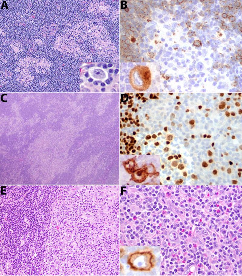
Figure 2. Histological features of type II CHL-RT.
A. Moth-eaten pattern, with foci of CHL in a background of CLL. Insert in A shows HRS cell. B. CD20 is weakly positive in CLL negative in HRS cell and surrounding T-cells. Inset shows CD30 staining of an HRS cell. C. Moth-eaten pattern, more extensive involvement. HRS cells are weakly positive for PAX5 in D, and are positive for CD15 as shown in inset. E. Segregated pattern. A distinct border is seen between CLL and CHL F. Typical histological features of CHL are present with mixed inflammatory background. Inset shows CD30 positivity of HRS cell.

