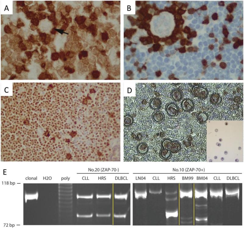
Figure 4. Microdissection and Clonal Analysis.
A. ZAP-70 positive CLL. Arrow indicates a HRS cell (ZAP-70 negative). T cells are strongly positive for ZAP-70. B. ZAP-70 negative CLL. ZAP-70 positive T cells rosette a HRS cell. C. PAX-5 and CD30 double staining. HRS cells are double positive and background CLL cells are PAX-5 positive but CD30 negative. D. Microdissection. Insert shows HRS cells after microdissection. E Clonal analysis of two concurrent CHL-RT and DLBCL-RT cases. DNA molecular markers with 118 bp and 72 bp shown; clonal, monoclonal positive control; poly, polyclonal control. Microdissected CLL, HRS and DLBCL cells were compared. Yellow lines indicate different sites. Left panel (ZAP-70− case): HRS and CLL cells microdissected from an inguinal lymph node, and DLBCL microdissected from bone marrow. Right panel (ZAP-70+ case): LN04, CLL, HRS from a supraclavicular lymph node in 2004; BM99 from bone marrow in 1999 when CLL was initially diagnosed; BM04, CLL and DLBCL from bone marrow in 2004.

