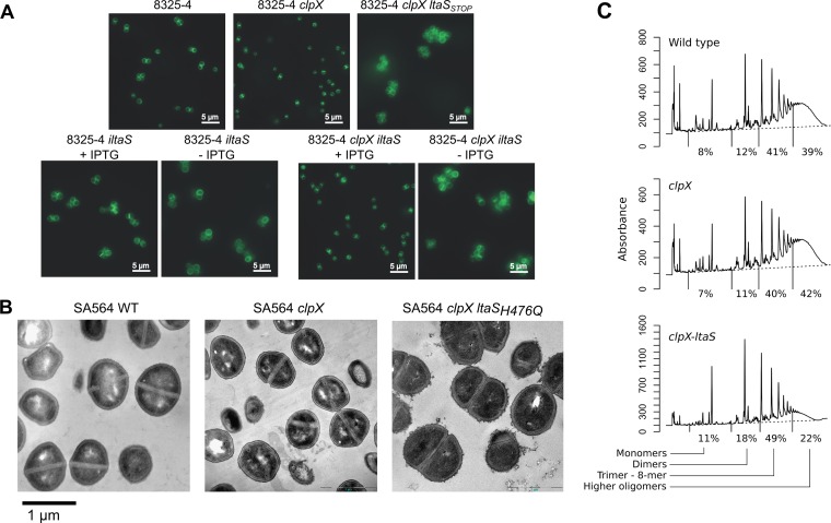
FIG 5 .
Cell morphology, septum formation, and cell wall structure are affected by inactivation of clpX and by the compensatory loss of LtaS activity. (A) Fluorescence microscopy analysis of wild-type, clpX-inactivated, and LTA-depleted strains. S. aureus strain 8325-4 and the corresponding clpX and clpX-ltaSSTOP mutants were grown in TSB to exponential phase at 37°C. Strains 8325-4-iltaS and 8325-4 clpX-iltaS were grown to exponential phase at 37°C in the presence of 1 mM IPTG and then diluted into medium with or without 1 mM IPTG and grown for 3 h at 37°C. Bacteria were stained with BODIPY-vancomycin and prepared for fluorescence microscopy as described in Materials and Methods. Examples of raw microscope images can be seen in Fig. S4 to S7 in the supplemental material. (B) TEM analysis of wild-type, clpX-inactivated, and LTA-negative strains. The SA564 wild type, the SA564 clpX strain, and the 564-25-2 (SA564 clpX ltaSH476Q) strain were grown in TSB to log phase at 37°C and prepared for electron microscopy as described in Materials and Methods. (C) Peptidoglycan structure in the SA564 wild type, the SA564 clpX strain, and the 564-25-2 (SA564 clpX ltaSH476Q) strain. The figure panel shows HPLC chromatograms of mutanolysin-digested peptidoglycan purified from the indicated strains and percentages indicating the relative abundances of monomers, dimers, trimers to 8-mers, and higher oligomers in each strain. Results representative of two individual experiments are shown.

