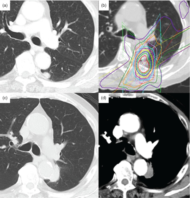
Fig. 1.
Chest CT findings. (a) Chest CT 3 years ago shows the lesion of nodule in left lower lobe. (b) Stereotactic radiotherapy treatment planning image. (c, d) Three years after SRT, an abnormal shadow appeared. PET-CT revealed soft tissue opacity and an irregular nodule 7 mm in size, leading to a diagnosis of local recurrence of lung cancer. CT: computed tomography; SRT: stereotactic radiotherapy; PET-CT: positron emission tomography-CT

