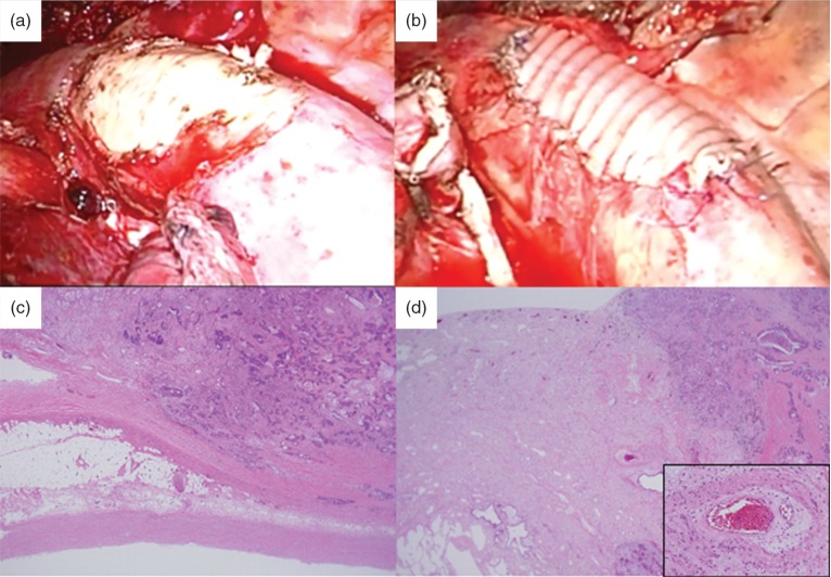
Fig. 3.
Surgical and pathological findings. (a) We performed combined aortic resection. Only a small amount of bleeding resulted from insertion of the stent graft. (b) The aortic wall defect was reinforced with a vascular graft patch. (c) Hematoxylin-Eosin stain shows fibrous hypertrophy and adhesions between the visceral pleura and the aorta. (d) An interstitial pneumonitis with cicatricial perivascular fibrosis can be seen. The small muscular arteries exhibit prominent myointimal proliferation and intramural hyalinization. There are pathological changes indicating chronic radiation fibrosis.

