Figure 2.
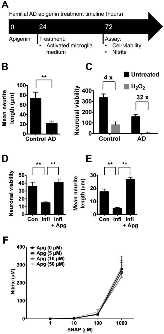
Apigenin treatment regime for familial AD iPSC-derived neurons (A). Neurons were generated from iPSCs from a familial AD patient carrying a PSEN1 (P117R) mutation or an age-matched control. (B) Length of neurites from familial AD or control neurons was measured using HCA-vision software. All neurites were measured in >10 images per experiment, n = 3. **indicates significant difference (p ≤ 0.01), paired t-test. (C) Neurons were treated with vehicle control or 100 μM H2O2 for 24 h and viability of AD or control neurons was measured. (D) Neuronal viability and (E) neurite length were measured in AD neurons or those cultured in media taken from activated microglia under inflammatory conditions (Infl; microglia activated with LPS (50 μg/ml) and IFN-γ (20 U/ml) for 48 h ± 50 μM Apigenin (Apg; 24 hour pre-incubation). (F) Nitrite formation in cell culture medium in the absence of cells treated with SNAP (0, 1, 10, 100, 1000 μM) and apigenin (0, 5, 10, 50 μM). Data shown are mean ± standard error of the mean from 3 independent experiments.

