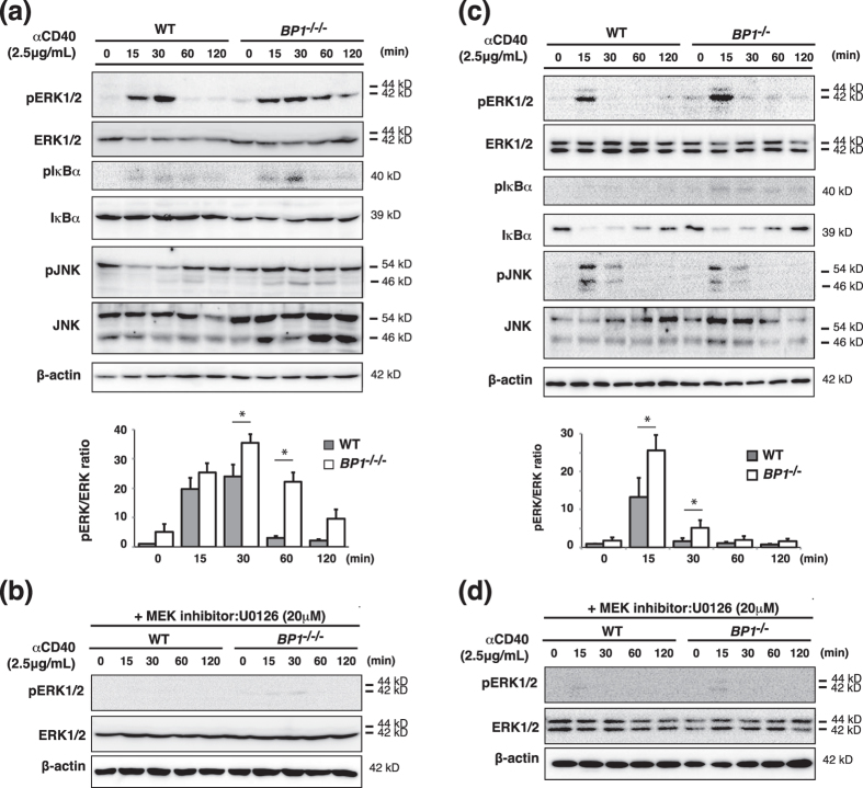
Figure 2. Disruption of TAX1BP1 in the DT40 B cell line enhanced ERK pathway activation in response to CD40 stimulation.
Western blot of whole cell lysates stimulated for the indicated time points with anti-CD40 antibody (αCD40). The results are representative immunoblot analyses from three independent experiments. The amount of ERK protein phosphorylation was determined as the ratio of phospho-ERK protein to total ERK protein and normalized with respect to unstimulated WT cells. The results are shown as the means and standard deviation (n = 3). Statistical analysis was performed by two-tailed Student’s t tests (*P < 0.05 compared with WT samples). (a) Whole cell lysates were prepared from αCD40 stimulated WT DT40 cells (WT) and TAX1BP1-deficient DT40 cells (BP1−/−/−). (b) The MEK inhibitor U0126 reduced ERK pathway activation in TAX1BP1-deficient cells. Shown is a western blot of whole cell lysates stimulated for the indicated time points with αCD40 in the presence of U0126 (20 μM). (c) Whole cell lysates were prepared from αCD40 stimulated splenic B cells from WT and TAX1BP1-deficient mice (BP1−/−). (d) The MEK inhibitor U0126 reduced ERK pathway activation in splenic B cells from TAX1BP1-deficient mice (BP1−/−). Shown is a western blot of whole cell lysates stimulated for the indicated time points with αCD40 in the presence of U0126 (20 μM).

