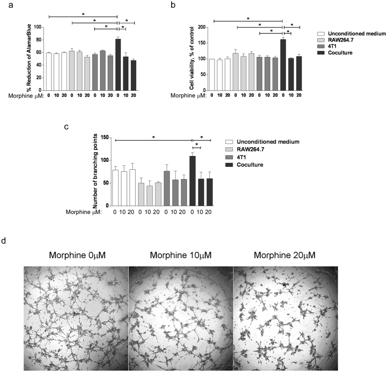
Figure 1. Morphine prevents the ability of CM from co-cultured cells to elicit EC proliferation and tube formation.
(a) Determination of BAEC proliferation using the AlamarBlue assay. BAEC were exposed for 48 h to the conditioned media of 4T1, RAW264.7 cells grown alone or together in the presence (10 or 20 μM as indicated) or absence of morphine. Cells were added with AlamarBlue Reagent and incubated for 4 h. The absorbance at 570 nm and 600 nm was recorded. Cell viability is presented as the percentage of AlamarBlue reagent reduction. Mean ± S.D. is shown for N = 3 independent experiments. *P = 0.05. (b) Determination of BAEC proliferation using the MTT assay. BAEC were exposed for 48 h to the conditioned media of 4T1, RAW264.7 cells grown alone or together in the presence (10 or 20 μM as indicated) or absence of morphine. Cells were incubated for 5 h in MTT-containing medium. The absorbance at 595 nm was determined. Cell viability is presented as the percentage of viability of control cells. Mean ± S.D. is shown for N = 3 independent experiments. (c) BAEC were incubated with either 4T1, RAW264.7 cell-conditioned media or unconditioned media as the control and added onto polymerized matrigel for 6 h. Capillary-like tubes were imaged for quantification. Results are reported as the mean number of branching points ± S.D. N = 3 independent experiments. *P = 0.05. (d) Representative images of the capillary-like tubes formed by BAEC.

