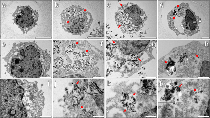
Figure 8.
Representative electron micrographs from TEM of Spurr resin-sectioned (100 nm sections) native THP-1 cells (a,e,i), THP-1 cells co-cultured with 50 μg/mL Alhydrogel® (24 h) (b,f,j) and 50 μg/mL Adju-Phos® (Brenntag Biosector, Denmark) adjuvant (24 h) (c,g,k) and 50 μg/mL Imject™ Alum (Pierce, Thermo Scientific) adjuvant (24 h) (d,h,l). Cell resin-sections were stained for 20 min with 2% ethanolic uranyl acetate, rinsed with 30% ethanol followed by ultrapure water and finally allowed 24 h drying time prior to analysis via TEM. Inserts show close-ups of intracellular adjuvant particles contained within vesicle-like structures and the red arrows highlight their presence within the respective cell images. Magnification and scale bars: (a–c) X 8 K, 5 μm, (d) X 10 K, 2 μm, (e) X 15 K, 2 μm, (f–h) X 30 K, 1 μm, (i) X 30 K, 1 μm and (j–l) X 60 K, 0.5 μm, respectively.

