Fig. 1.

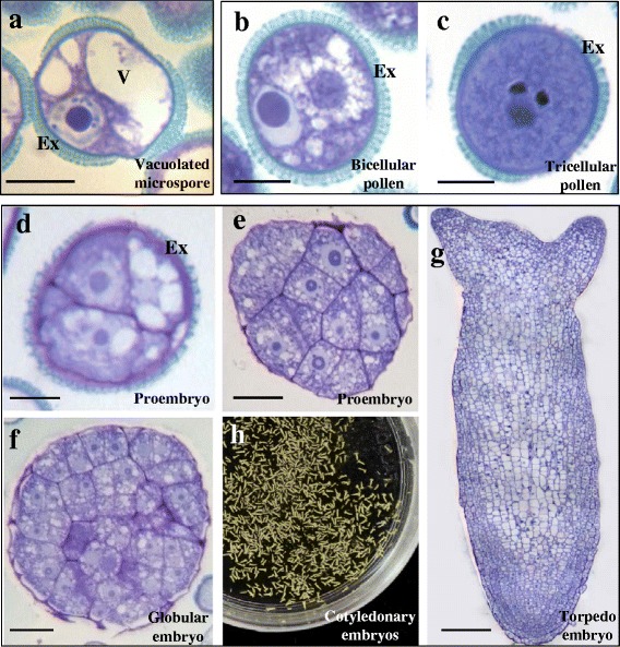
Main stages of the two microspore pathways: gametophytic development and microspore embryogenesis. a-g Semithin sections, toluidine blue staining. a Vacuolated microspore. b-c Gametophytic development, in vivo. b Bicellular pollen. c Tricellular mature pollen. d-h Microspore embryogenesis, in vitro. d Two-cell proembryo. e Multicellular proembryo. f Globular embryo. g Late torpedo embryo. h Panoramic view of cotyledonary embryos in the Petri dish. Ex: exine, V: vacuole. Bars: a-d, 10 μm; e, f, 20 μm; g, 50 μm
