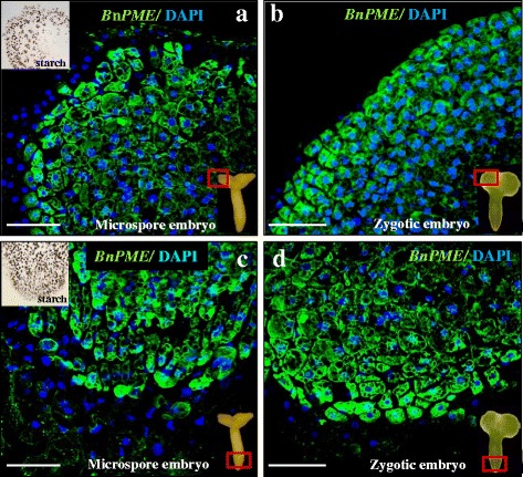
Fig. 5.

In situ PME expression by FISH in cotyledonary microspore embryos and zygotic embryos. Confocal merged images of fluorescence provided by DAPI staining of nuclei (in blue) and fluorescent in situ hybridization signal (in green). a, c Microspore embryo. b, d Zygotic embryo. a, b Cotyledon region of embryos, as indicated by the square. c, d Radicular region of embryos, as indicated by the square. Insets in a and c: Iodide specific staining for starch, analogous cotyledonar and radicular embryo regions as in “a” and “c”. Bars: 50 μm
