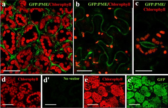
Fig. 6.

In vivo GFP: PME subcellular localization in transformed tobacco leaves. Confocal images of fluorescence provided by chlorophyll autofluorescence (in red) and GFP fluorescence (in green). a, b, c GFP: PME transformed leaves, merged images of red and green channels of mesophyll a, pavement epidermal b and stomatal guard e cells. d, d’ Wild type leaf. e, e’ GFP transformed leaf. d, e Red channel. d’, e’ Green channel. Bars: 25 μm
