Fig. 8.

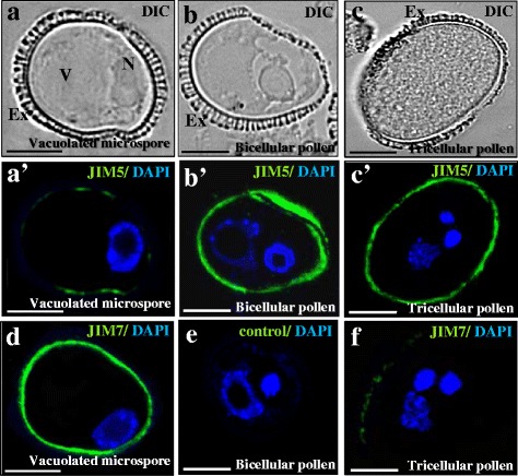
Immunolocalization of esterified and non-esterified pectins during gametophytic development. a, b, c are the same structures, visualized by DIC (Differential Interference Contrast, Nomarsky) as in a’, b’, c’. a’, b’, c’ Immunofluorescence of esterified (JIM7 antibody) and non-esterified (JIM5 antibody) pectins during gametophytic development. a, a’, d Vacuolated microspore; b, b’, e Bicellular pollen; c, c’, f Tricellular mature pollen. a’, b’, c’ Merged images of fluorescence provided by DAPI staining of nuclei (in blue) and JIM5 immunofluorescence signal (in green). d, f Merged images of fluorescence provided by DAPI staining of nuclei (in blue) and JIM7 immunofluorescence signal (in green). e Control immunofluorescence experiment avoiding the first antibody. Ex: exine, N: nucleus, V: vacuole. Bars: 10 μm
