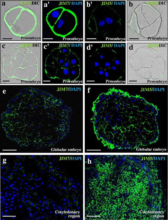
Fig. 9.

Immunolocalization of esterified and non-esterified pectins during microspore embryogenesis. Confocal merged images of immunofluorescence signal (green) and DAPI staining of nuclei (blue). For some stages, DIC image (Differential Interference Contrast, Nomarsky) of the same section is shown to reveal the structure (left side for each pair of images). a, a’, c, c’, e, g JIM7 immunofluorescence. b, b’, d, d’, f, h) JIM5 immunofluorescence. a-d Two-cell and early proembryos. e, f Globular embryos. g, h Cotyledonary embryos. Bars: a-d) 10 μm; e-h) 25 μm
