Fig. 1.

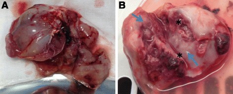
VX2 tumor liver development. a Picture reveals resected hepatic tumor after 4 week from VX2 tumor pieces implant, b Tumor bisected shows necrotic core (asterisk) and peripheral viable tumor (arrows)

VX2 tumor liver development. a Picture reveals resected hepatic tumor after 4 week from VX2 tumor pieces implant, b Tumor bisected shows necrotic core (asterisk) and peripheral viable tumor (arrows)