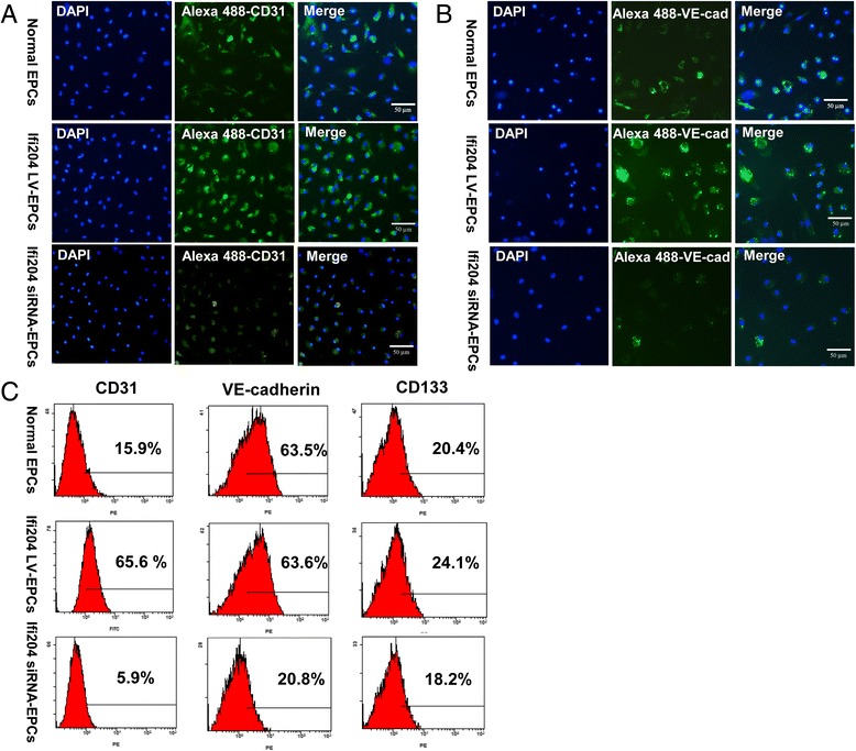
Fig. 3.

Assessment of endothelial markers in cultured EPCs by immunocytostaining and FACS. Cells were cultured and induced with Ifi204 lentivirus or Ifi204 siRNA. Adherent cells were stained with endothelial marker CD31 a and VE-cadherin b. Bar, 50 μm. c Cell surface markers investigated by FACS were as follows: endothelial cell (CD31 and VE-cadherin) and stem cell (CD133). Cellular surface markers were analyzed on normal EPCs (upper panel), Ifi204 lentivirus-transduced EPCs (middle panel), and Ifi204 siRNA-transfected EPCs (lower panel). Percentages of each marker indicated in the histograms (n = 3). EPC endothelial progenitor cell
