Fig. 8.

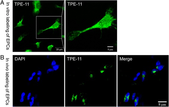
In-vitro and in-vivo staining of EPCs by the nanofluorogen. a EPCs were incubated with TPE-11 for 12 hours. Fluorescent images of the cells observed by a confocal microscope at the 405 nm excitations. Bar, 20 μm and 8 μm. b Sections of the ischemic gastrocnemius muscle were observed by a confocal microscope at the 405 nm excitations. Bar, 8 μm. EPC endothelial progenitor cell
