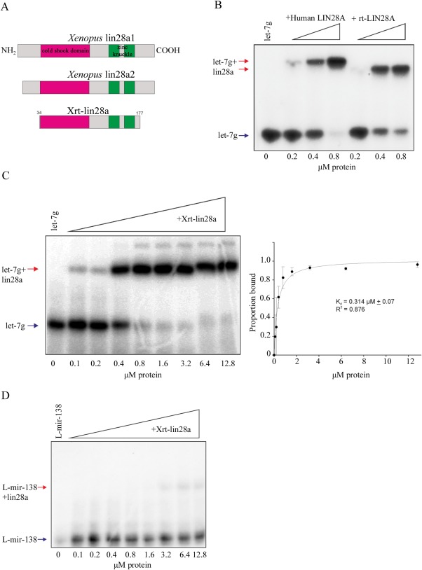
Figure 3.

A: Scale diagram of the Xenopus proteins used in this study. Cold shock domains are shaded magenta and zinc knuckles green. B: EMSA performed with 32P‐labelled L‐let‐7g and indicated concentrations of human recombinant LIN28A protein, either full‐length or truncated (rt). Arrows indicate labelled RNA (blue) and LIN28A‐RNA complex (red). C: EMSA performed with 32P‐labelled L‐let‐7g and indicated concentrations of Xrt‐lin28a. Gel shown is representative of n = 3. Arrows indicate labelled RNA (blue) and lin28‐RNA complex (red). Band intensities were quantified from three independent experiments and the proportion bound was calculated. Data were fit by nonlinear regression as described in Materials and Methods. Bmax = 1.017. D: EMSA performed with 32P‐labelled L‐mir‐138 and indicated concentrations of Xrt‐lin28a. Arrows indicate RNA and lin28a‐RNA complex. Gel shown is representative of n = 3. Arrows indicate labelled RNA (blue) and lin28‐RNA complex (red).
