
Figure 4.
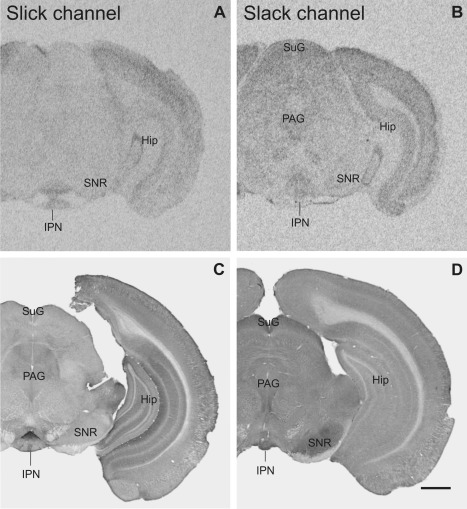
Overview of Slick and Slack channel distribution in the mouse brain (Bregma approximately –3.3 mm). A,B: Strong mRNA labeling was detected in in situ hybridization in the interpeduncular nucleus for Slick and Slack channels, respectively. B: Slack channel mRNA signal was moderate to strong in the superficial gray and in periaqueductal gray. C,D: The interpeduncular nucleus exhibited high immunoreactivity for both channels. D: Slack channel immunoreactivity was high in the substantia nigra pars reticulata, C whereas Slick channel immunostaining was only weak. Scale bar = 1,000 μm.