
Figure 5.
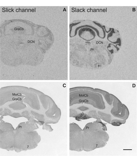
Slick and Slack channel mRNA expression and immunoreactivity in the mouse brain (Bregma approximately –6.2 mm). A,C: In the cerebellar cortex, Slick channel showed only low signals in in situ hybridization and immunohistochemical experiments. B: In contrast, we detected particularly strong signals for Slack channel mRNA in granule cells and in the Purkinje cell layer. C: Slick channel immunohistochemistry demonstrated only weak staining in cerebellar cortex. D: Slack channel immunostaining was moderate in the granule cell layer and only very weak staining of Purkinje cell bodies. However, strong immunolabeling was detected in the molecular layer of the cerebellar cortex. Scale bar = 1,000 μm.