
Figure 9.
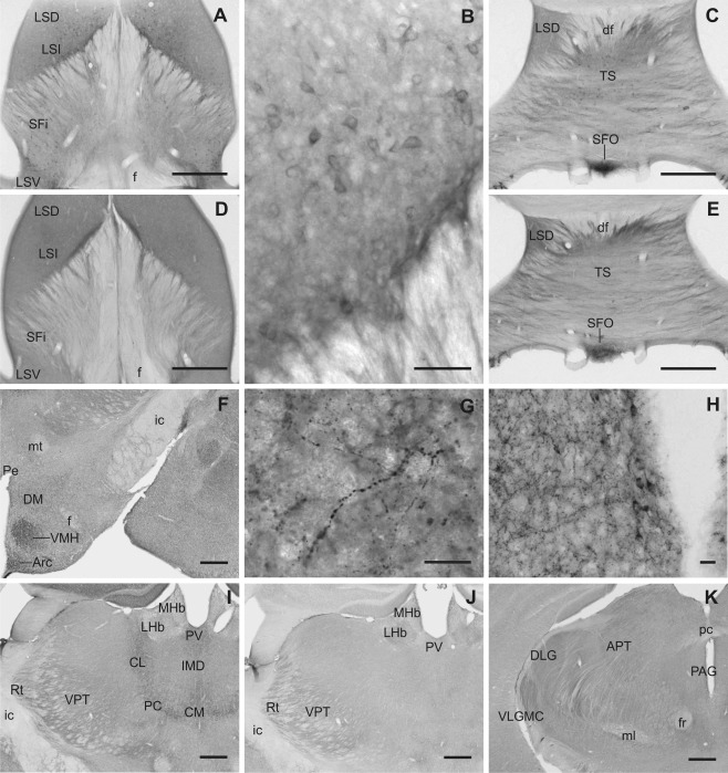
Slick and Slack channel immunoreactivity in septal nuclei, thalamus, and hypothalamic areas. A,B: Intense Slick channel immunolabeling of cell bodies was observed in the intermediate part of lateral septal nucleus (Bregma approximately 0.0 mm). D: Slack channels showed a diffuse moderate to strong immunostaining in all parts of lateral septal nuclei (Bregma approximately 0.0 mm). Slick (C) and Slack (E) channel immunolabeling showed high signals in cell bodies of the subfornical organ (Bregma approximately –4.5 mm). F–H: Slick channel immunoreactivity in hypothalamic areas. F: Prominent labeling of numerous processes and varicosities was found in the ventromedial hypothalamic and in the arcuate nucleus. In the dorsomedial hypothalamic nucleus, overall Slick channel immunoreactivity was only moderate; nevertheless, intense staining of processes and varicosities could be observed, as shown in G (Bregma approximately –1.6 mm). H: For the periventricular nucleus, we observed intense Slick channel staining of processes and varicosities. Strong staining of cell bodies and their proximal processes was observed throughout the periventricular nucleus but was most striking in ventral parts (Bregma approximately –1.6 mm). I: Slick channel immunoreactivity was observed in cell bodies of the centromedial, paracentral, and centrolateral thalamic nuclei. The reticular and ventral posterior thalamic nuclei showed a diffuse staining for the Slick channel (Bregma approximately –1.7 mm). J: Lateral habenula, reticular thalamic nucleus, and ventral posterior thalamic nuclei were immunoreactive for the Slack channel (Bregma approximately –1.7 mm). K: Most intense Slack channel immunoreactivity within the thalamus was observed in the magnocellular part of the ventrolateral geniculate nucleus. Moderate staining was also detected in dorsal lateral geniculate and in anterior pretectal nuclei (Bregma approximately –2.8 mm). Scale bars = 500 μm in A,C–F,I–K; 50 μm in B; 20 μm in G,H.