Figure 4.

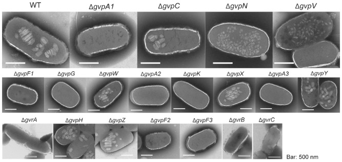
Transmission electron microscopy images of wild type (WT) and several gvp mutants. WT cells and mutants were grown in LB in sealed universals for 24 h and gas vesicles were observed by TEM (scale bars represent 500 nm).

Transmission electron microscopy images of wild type (WT) and several gvp mutants. WT cells and mutants were grown in LB in sealed universals for 24 h and gas vesicles were observed by TEM (scale bars represent 500 nm).