
Figure 1.
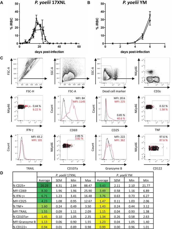
Early NK‐cell activation during lethal and nonlethal P. yoelii infection. C57BL/6 mice were infected i.p. with 105 RBCs infected with (A) P. yoelii 17XNL or (B) P. yoelii YM. Each line represents the mean (SEM) parasitemia for groups of three to five mice in each of three (A) or two (B) independent experiments. (C) Splenic NK cells were identified by flow cytometry. Lymphocytes were gated after exclusion of cell aggregates, followed by exclusion of dead cells. NK cells were identified as NKp46+ CD3ε– lymphocytes. Overlays are shown for activation and functional markers. Black = naive animal; red = day 1 of P. yoelii 17XNL infection. (D) Changes (fold increase) in frequency (%) or expression levels (MFI) of activation and functional markers on murine splenic NK cells 24 h after infection with P. yoelii 17XNL or P. yoelii YM compared to naive controls were determined by flow cytometry. Data are shown as mean, SEM, and range for P. yoelii 17XNL (n = 13 mice) and P. yoelii YM (n = 10 mice) infection pooled from three (Py17XNL) or two (PyYM) independent experiments.