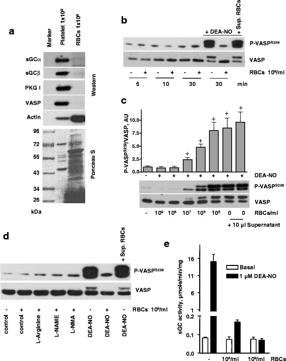
Fig. 3.

Inhibition of sGC activity by RBCs. a Western blot analysis of sGC, PKG, VASP, and actin expression in platelet and RBC lysates. Upper panel represent Western blot and lower panel represent Ponceau-S-stained membrane. b-d) Western blot analysis of VASPS239 phosphorylation in platelets (3x10⁸/ml) treated with 1 μM DEA-NO in the presence of RBCs (5x10⁸/ml, in 10 μl). b Platelets were coincubated with RBCs (109/ml, 10 μl), or the same volume (10 μl) of supernatant from the last wash of RBCs for indicated time. When indicated, samples were treated with DEA-NO (1 μM, 30 min). c Indicated amount of RBCs (10 μl) were added to platelet suspension for 10 min, and then stimulated with DEA-NO (1 μM, 1 min). When indicated, the same volume (10 μl) of supernatant from the last wash of RBCs was added to the platelets. For the bar graphs, immunoblots were scanned and quantified by the Image J program. The intensity of the VASPSer239 signal was normalized to the total VASP signal, which was designated as 1 in control samples. Results are means ± SEM, n = 4, + significant difference from control samples. d Platelets (3x10⁸/ml) were coincubated with RBC (5x10⁸/ml, 10 μl) and treated with L-Arginine, L-NAME, or L-NMA for 10 min. When indicated, samples were treated with DEA-NO (10 μM, 1 min). Total VASP blots served as loading control. Shown are representative blots of three independent experiments. e Basal and NO-induced (1 μM DEA-NO) purified sGC activity measured in anaerobic conditions after incubation with buffer or indicated amounts of RBC. Data are presented as mean ± SEM, n = 4
