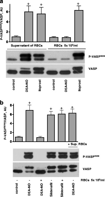
Fig. 4.

RBCs inhibits platelet PKG stimulated by NO donors, but not by PDE5 inhibition. a, b Western blot analysis of VASPS239 phosphorylation in platelets (3x10⁸/ml) stimulated by DEA-NO (1 μM, 1 min), iloprost (2 nm, 1 min) or sildenafil (1 μM, 5 min) in the presence of washed RBCs (1x 10⁸, 10 μl) or supernatant (10 μl) from last wash of RBCs. a Supernatant from RBC (10 μl) washed by HEPES buffer, or RBCs (1x10⁸/ml, 10 μl) were added to platelets for 10 min, then stimulated with DEA-NO, or iloprost. b RBCs (108/ml, 10 μl), or supernatant from RBC (10 μl) were added to platelets for 10 min, then stimulated with DEA-NO, or with sildenafil. Total VASP blots served as loading control. Shown are representative blots of 4 independent experiments. For the bar graphs, immunoblots were scanned and quantified by the Image J program. The intensity of the VASPSer239 signal was normalized to the total VASP signal, which was designated as 1 in control samples. Results are means ± SEM, n = 4, + significant difference from control samples
