Figure 3.


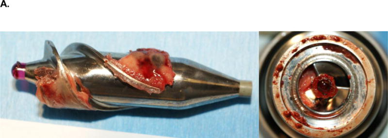
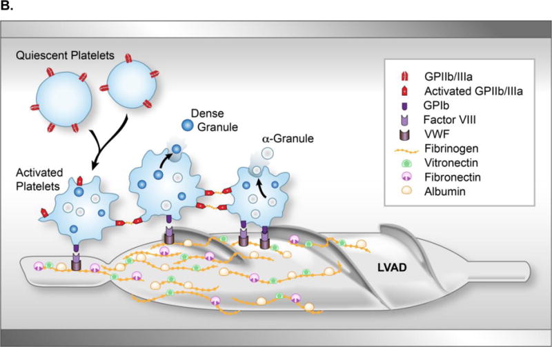
Clot formation on an axial flow pump. (A) Explanted HeartMate II LVAD with evidence of red- and white-clot around inflow as well as in main rotor. (B) Upon contact with the blood, the titanium bioreactive surface of the LVAD adsorbs several plasma proteins, including albumin, fibrinogen, fibronectin and vitronectin forming a protein layer covering the LVAD surface. With contact, activated platelets change their discoid shape by putting out pseudopods and release their dense and α-granules. Fibrinogen, vWF, and factor VIII form a complex with the GP1b receptor site of the activated platelet to cause adherence. This complex produces a conformational change in the GPIIb/IIIa receptor allowing the platelet to bind with fibrinogen. As this occurs, the newly recruited platelets release their granule content, which accelerate the thrombus development by increasing linkages between the platelets with fibrinogen/fibrin bridges. Subsequently thrombin is generated and stabilizes the local thrombus. (vWF, von Willebrand factor; GP, glycoprotein; LVAD, left ventricular assist device)
