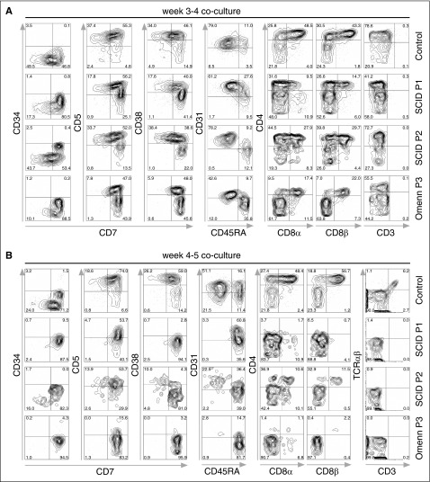
Figure 4.
In vitro T-lineage differentiation of control and SCID iPSC lines. Flow cytometric analysis of T-lineage developmental progression of control- and patient-derived cells. iPSCS were allowed to differentiate for 8 days into embryoid bodies, and magnetic bead-purified CD34+ cells were cocultured with OP9-DL-4 cells. (A) Cells from P1 and P2 with SCID, and from P3 with OS attained normal expression of early markers of T-lineage differentiation (CD7, CD5, and CD38) upon 3 to 4 weeks of coculture with OP9-DL-4 cells. (B) After 4 to 5 weeks of coculture, cells from a healthy control progress to the CD4+ CD8αβ+ DP stage of differentiation, with the appearance of CD3+ TRA/TRB+ cells. By contrast, SCID- and OS-derived cells were mostly blocked at the CD7+ CD31−/+ CD45RA+ stage of differentiation, with a virtual absence of CD4 and CD8α/β expression, and lack of CD3+ cells. In (A-B), cells were pre-gated for lymphocytes (SSCxFSC), DAPI-, and CD45+. DAPI, 4′,6-diamidino-2-phenylindole; FSC, forward scatter; SSC, side scatter.

