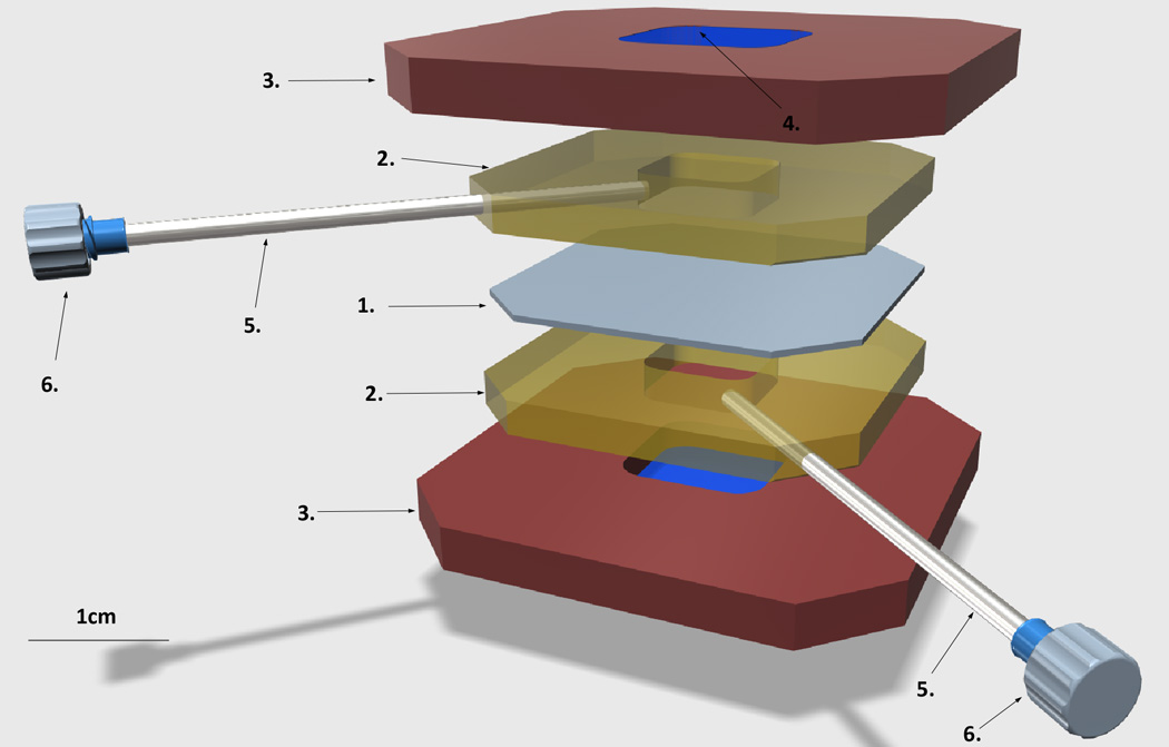
Figure 1. Diagram of the liposome dialyzer.

1. Dialysis membrane, 20K MWCO
2. Silicone gaskets
3. Plastic cassette, top and bottom
4. The window on either side of the plastic cassette, sealed with the PCR film
5. Needles for loading sample and wash buffers
6. Needle luer caps
Sample chambers are colored yellow.
