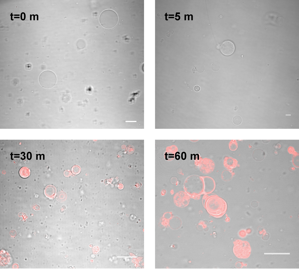
Figure 5. Staining of giant phospholipid vesicles.
Overlay of the confocal fluorescence and DIC bright field images of POPC vesicles incubated with Vybrant DiI Cell labeling solution for the specified time intervals. Scale bars are 15 µm.
The methods for preparation of liposomes were described in our previous work.1,21

