
Figure 6.
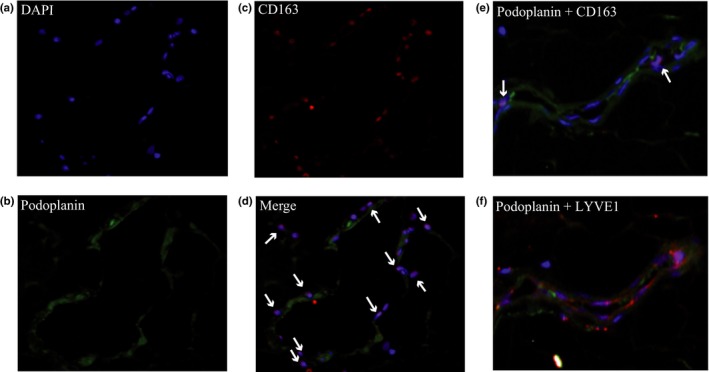
Immunofluorescent staining for intranodal macrophages adhered to lymphatic vessels of human metastatic lymph nodes. Nuclei of cells in metastatic lymph nodes were stained with DAPI (a). Images of lymphatic vessels stained with anti‐podoplanin antibody (green) (b), macrophages stained with anti‐CD163 antibody (red) (c), and merged images (d) are shown. White arrows indicate macrophages surrounding and contacting lymphatic vessels. (e) Merged images of the lymphatic vessels stained with anti‐podoplanin antibody (green) and anti‐CD163 antibody (red) are shown. White arrows indicate macrophages. (f) The same section was stained with anti‐podoplanin (green) and anti‐lymphatic vessel endothelial hyaluronan receptor (LYVE)1 antibody (red). Podoplanin‐positive cells are coexpressing LYVE1.