Figure 1.

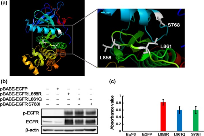
Crystal structure and oncogenic activities of EGFR L861Q and S768I mutations. (a) Structure of epidermal growth factor receptor (EGFR). The figures were drawn using the PyMOL Molecular Graphics System based on the crystal structure information from PDB ID 4G5J. Codon 768 is located in the alpha‐C helix, and both codons 858 and 861 are located in the activation loop. (b) Expressions of EGFR in the transfectant Ba/F3 cell lines. EGFR overexpression was confirmed by a western blot analysis. EGFR was strongly expressed in the Ba/F3 cell lines harboring each EGFR mutation. The phosphorylation level of EGFR was also elevated in the Ba/F3‐L861Q and Ba/F3‐S768I cell lines, similar to that in the Ba/F3‐L858R cell line. β‐actin was used as an internal control. p‐EGFR, phospho‐EGFR. (c) IL‐3 independent cell growth assay. A cell growth assay without IL‐3 was performed using an MTT assay. The Ba/F3 and Ba/F3‐EGFP cell lines could not grow in the absence of IL‐3, while all the Ba/F3 cell lines harboring each EGFR mutation could grow IL‐3 independently. Columns, mean of independent triplicate experiments; error bars, SD.
