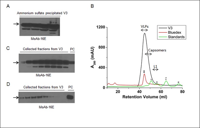
Fig 4. Ammonium sulfate precipitation and purification of the V3 infiltrated plants.
(A) Immunoblot of ammonium sulfate precipitated L1 protein samples collected at various stages of purification. 16E monoclonal antibody was used to detect the target L1 protein. (B) The size exclusion chromatogram for purification of ammonium sulfate precipitated L1 protein and VLPs using FPLC system. The running profile of molecular standards, BD; bluedex (2000 kDa), C; conalbumin (75 kDa), O; ovalbumin (43 kDa), CA; carbonic anhydrase (29 kDa) and R; ribonuclease (6.5 kDa) are showed on the chromatogram. (C) Immunoblot showing the detection profile of fig B and L1 protein was detected in the collected fractions corresponding to 40–50 mL fraction size. (D) Immunoblot showing the bands of cation exchange chromatography collected L1 protein. Sample (V3) and purified VLPs from insect cells (PC) were eluted at 0.3 M and ≥ 0.7 M NaCl, respectively. The arrows in immunoblots show HPV L1 band at 56 kDa.

