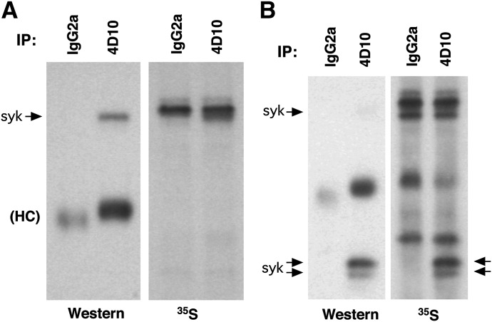
Figure 1. Metabolic labeling of cellular proteins in basophils.
Purified human basophils were incubated for 24 h with 5 ng/ml IL-3, and the cells were pulsed with [35S]Met for 18 h and then lysed for IP with IgG2a or 4D10 (subclass, IgG2a). Immunoblot (Western) or radiography was performed. (A) Lysis of the cells including a standard mix of proteolytic inhibitors. (B) Lysis of cells excluding proteolytic inhibitors.

