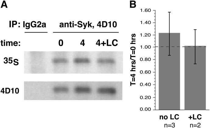
Figure 2. Chase component of the [35S]Met labeling of basophils.
Purified human basophils were incubated for 24 h with 5 ng/ml, and the cells were pulsed with [35S]Met for 18 h, washed, and placed back into similar medium without [35S]Met. In 2 experiments, conditions included ±100 μM lactacystin A. Samples at 0 and 4 h were removed and lysed for IP. (A) An example of the Western blot and autoradiographs. (B) Average of the experiments.

