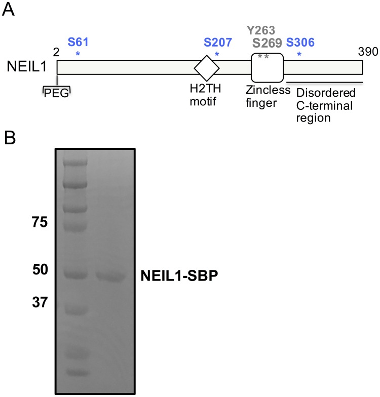
Fig 1. Sites of phosphorylation within the NEIL1 DNA glycosylase.
(A) Domain map of NEIL1 indicating the position of known sites of phosphorylation. The residues S207, S306, and S61 identified in this study are shown in blue and the Y263 and S269 sites previously identified [39] are indicated in black. (B) SDS-PAGE gel of SBP-tagged NEIL1 after affinity pull-down from HEK293T cell-extracts overexpressing NEIL1. The gel was stained with Coomassie blue and the NEIL1-SBP band was cut from the gel and digested with trypsin for identification of phosphorylated peptides via LC-MS/MS.

