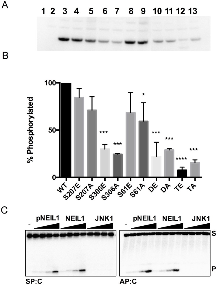
Fig 6. Phosphorylation of NEIL1 by JNK1 using an in vitro kinase assay.
(A) In vitro kinase assays were performed with NEIL1 constructs expressed from E. coli cells and purified to homogeneity and active JNK1 kinase for 30 minutes at 32°C. γ-32P incorporation was quantified using phosphor-autoradiography after SDS-PAGE analysis of the samples. Lane 1, no JNK1 control; lane 2, no NEIL1 control; lane 3–13 are WT, S207E, S207A, S306E, S306A, S61E, S61A, DE, DA, TE, TA, respectively. (B) Graphical representation of two experimental repeats of the in vitro kinase assay. Statistically significant values (at 95% confidence) were determined by a one-way Anova test where * denotes p-values <0.05, *** denotes p-values <0.0005, and **** denotes p values <0.0001. (C) Glycosylase activity assays were performed using Sp:C and AP:C substrates with increasing amounts of in vitro phosphorylated NEIL1 (pNEIL1), unphosphorylated NEIL1 (positive control), and JNK1 (negative control). S and P indicate substrate and product, respectively.

