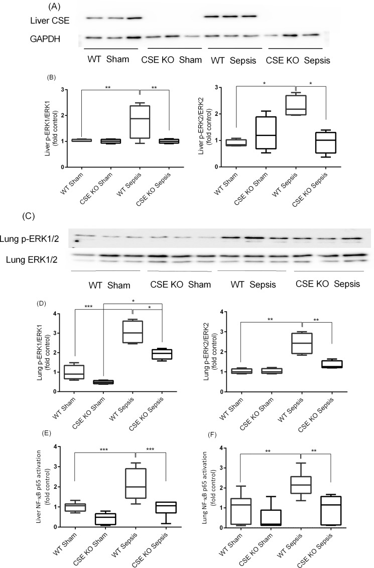
Fig 4. Effect of CSE Gene Deletion on the Phosphorylation of ERK1/2 and NF-kB p65 Activation in the Liver and Lung Following CLP Induced Sepsis.
(A-B) Liver p-ERK1/2 expression. Phosphorylation of ERK1/2 (p-ERK1: P<0.01; p-ERK2: P<0.05) was increased following CLP induced sepsis in WT mice compared to sham operation controls. In CSE KO mice ERK1/2 phosphorylation (p-ERK1: P<0.01; p-ERK2: P<0.05) was reduced significantly following CLP induced sepsis compared to WT sepsis mice. (C-D) Lung p-ERK1/2 expression. Phosphorylation of ERK1/2 was increased (p-ERK1: P<0.001; p-ERK2: P<0.01) following CLP induced sepsis in WT mice compared to sham control. In CSE KO mice ERK1/2 phosphorylation was reduced significantly (p-ERK1: P<0.05; p-ERK2: P<0.01) following CLP induced sepsis compared to WT sepsis mice. Results were normalized with GAPDH and expressed as the relative fold increase of pERK1/2 expression compared with sham control. For western blot results, each lane represents a separate animal. The blots shown were representative of all animals in each group with similar results. (E-F) Liver and lung NF-κB p65 activation. Activation of NF-κB p65 (P<0.001) was increased following CLP induced sepsis in WT mice compared to sham control. In CSE KO mice NF-κB p65 activation (P<0.001) was decreased significantly following CLP induced sepsis compared to WT sepsis mice. Results were expressed as fold increase over control. Data represent the mean±standard deviation (n = 8). Data were analysed for Gaussian or Normal distribution using Shapiro-Wilk test. One-way ANOVA with post hoc Tukey’s test was performed to compare multiple groups. Statistical significance was assigned as *P<0.05; **P<0.01: and ***P<0.001.

