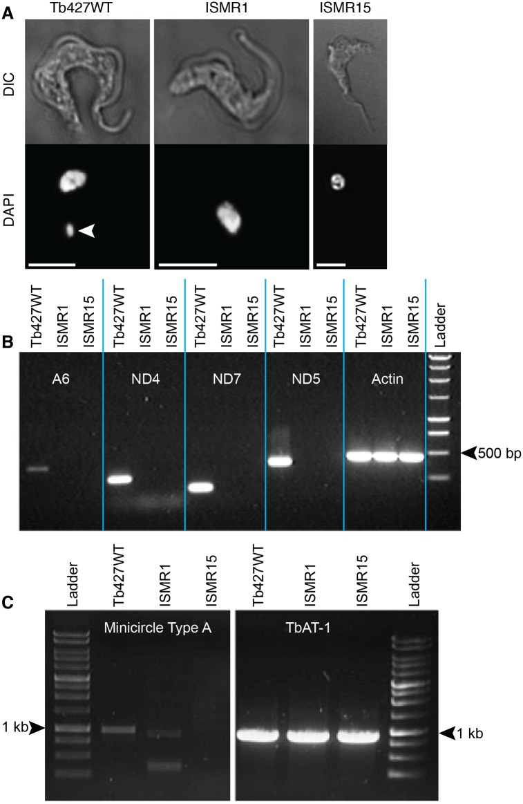
Fig 3. ISM resistant cell lines have lost their kinetoplasts.
(A) Fluorescence microscopy of DAPI-stained Tb427WT and ISMR1 and ISMR15 cell lines. The white arrowhead indicates the kinetoplast, which is absent in ISMR1 and ISMR15. The white scale bar represents a length of 5 μm. (B) Electrophoresis gel of PCR products of kinetoplast-encoded genes and nuclearly encoded actin. Genomic DNA was extracted from the parental Tb427WT strain as well as from the ISM resistant ISMR1 and ISMR15 strains and subjected to PCR amplification using primers specific for the gene fragments stated (S1 Table). (C) Like frame B but using primers specific for minicircles, using the TbAT1 gene as a positive control for a nuclearly-encoded single copy gene [62].

