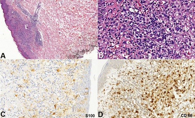
Figure 2. Histology and Immunostains of the skin biopsy. A - Dense infiltrate in the dermis (H&E, 100X); B - Infiltrate composed by histiocytoid cells, lymphocytes, plasma cells and multinucleated cells (H&E, 400X); C - S100 partially positive in epidermal Langerhans cells and dermal infiltrate (anti-S100, 200X); D - CD68 positive in histiocytoid cells of the dermal infiltrate (anti-CD68, 200X).

