
Figure 1. In vivo imaging and postmortem histopathology of choroid plexus.
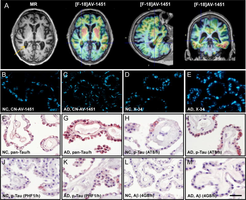
(A) MR and [F-18]AV-1451 PET images of a 91-year old AD patient. Choroid plexus is evident in the MR image (arrow) and shows high [F-18]AV-1451 retention in PET scans as denoted by crosshairs. (B–M) Fluorescence and light microscopy micrographs of choroid plexus from postmortem brains of a 83-year old NC and a 77-year old AD patient, stained with CN-AV-1451 (B,C), X-34 (D,E), total and phosphorylated tau (F–K) or Aβ immunohistochemistry (L,M). B–E: In both NC and AD choroid plexus, CN-AV-1451 and X-34 label tangles which contain fibrillar β-sheet structure. F–M: In NC and more prominently in AD, epithelial cells of the choroid plexus contain immunoreactivity (brown color) with antibodies against pan-Tau (F,G) and p-Tau antibodies AT8 (H,I) and PHF1 (J,K), but only light or no Aβ immunoreactivity (L,M, 4G8 antibody). Sections in F–M were counter-stained with hematoxylin (h, blue color). Scale bar = 35 μm (B–M).