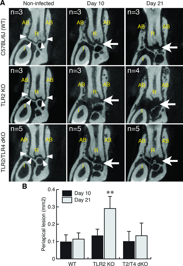
Figure 1. TLR2 KO mice exhibited extended periapical bone destruction compared to wild-type C57BL/6 and TLR2/TLR4 dKO mice.
A. Representative µCT images of periapical lesions in anterior-posterior direction. The effect of TLR2 and TLR2/TLR4 double deficiencies on periapical bone destruction was determined on days 10 and 21 post endodontic infection. Non-infected mice in each strain served as corresponding background controls. The sample number (n) of each group was indicated in each panel. The position of each CT slice was the most central part of mandibular first molar distal root (R). AB: alveolar bone. Arrowheads in the panel of non-infected controls indicate the margin of alveolar bone in periapical region. Arrow points to enlarged radiolucent area indicating the development of periapical lesion. The original length of the side of each square CT image was 2.4 mm.
B. Size of periapical lesions. The extent of periapical lesions was measured as previously described (AlShwaimi et al., 2013). Although the periapical lesion size on day 10 post-infection was similar in all genotypes, TLR2 KO mice exhibited significantly enlarged periapical bone loss compared to wild-type C57BL/6J and TLR2/TLR4 dKO mice on day 21. **: p<0.01 vs. either C57BL/6J or dKO, vertical bar: standard deviation.

