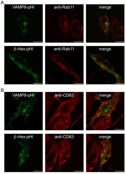
Figure 1. pHluorin-based exocytosis reporters are localized in Rab11-positive endosomes and lysosomes.
RBL-2H3 cells were transfected with VAMP8-pHl or β-Hex-pHl, fixed, permeabilized, probed with anti-Rab11 (A) or anti-CD63 (B) antibodies, and imaged by confocal microscopy. Scale bars are 10 μm. Representative images are shown.

