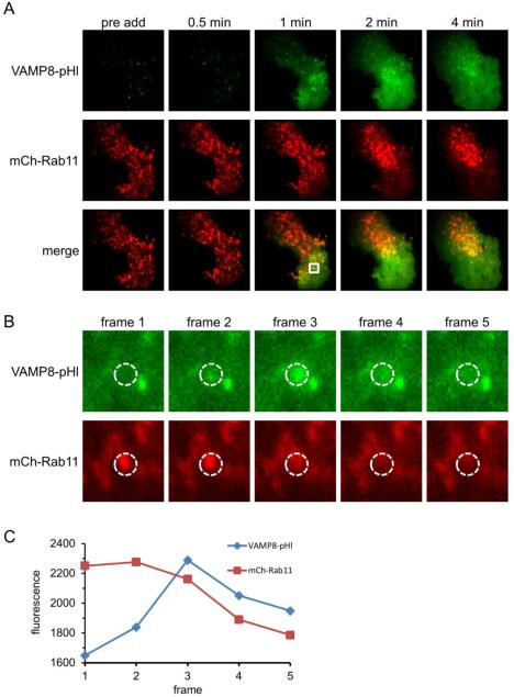
Figure 3. VAMP8-pHl exocytic events are frequently marked by Rab11.
RBL-2H3 cells co-transfected with VAMP8-pHl (green) and mCh-Rab11 (red) were visualized by two-color TIRF microscopy and stimulated with 200 ng/ml Ag. (A) Individual frames show a cell before Ag addition (pre add) and at various times after Ag addition. (B) Individual frames at 1 min post-Ag addition highlighting a mCh-Rab11-labeled vesicle fusing with the plasma membrane, resulting in the dequenching of vesicular VAMP8-pHl fluorescence. Frame 3 is same as 1 min frame from (A). Frame rate is 5.4 fps. (C) Integrated fluorescence traces of the region of interest (dashed circles) shown in (B) to quantify coordinated loss of mCh-Rab11 fluorescence and VAMP8-pHl fluorescence dequenching. Image boxes are 25.6 μm2 in (A) and 3 μm2 in (B). Scale bars are 5 μm in (A) and 0.6 μm in (B).

