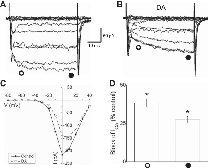
Fig. 3.
Dopamine reduced Ca channel currents in isolated mouse horizontal cells identified by tdTomato fluorescence. A: Ca channel currents recorded under voltage clamp in response to the 40-ms voltage step paradigm shown in Fig. 1C before dopamine application. B: Ca channel currents recorded during superfusion with 10 μM dopamine (DA) show reduction of inward current amplitude and slowed activation. C: steady-state current-voltage (I-V) relations measured in control conditions and in the presence of DA near the end of each voltage step (time of measurement denoted by filled circle below the current traces) show reduction of the current between −30 and +30 mV. D: % of peak control current that was blocked by DA ∼10 ms after beginning of each voltage step (open circle) and at end of voltage steps (filled circle). *P < 0.05.

