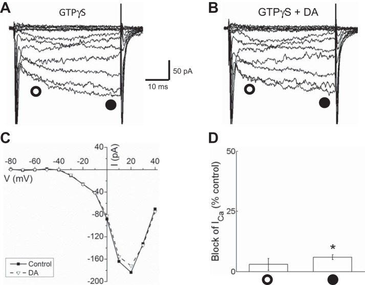
Fig. 9.
Activation of G proteins with GTPγS occluded inhibition of Ca channel currents by dopamine in isolated mouse horizontal cells. A: ensemble of traces recorded in the presence of GTPγS in the intracellular solution showing slowed activation kinetics. B: Ca channel currents in the same GTPγS-filled cell recorded after treatment with 10 μM dopamine showed modest inhibition. C: I-V relationships of Ca channel currents measured near the end of each voltage step (filled circles) from the same cell in A and B recorded from with GTPγS in the intracellular solution before (control) and after treatment with 10 μM dopamine (DA). D: % of peak control current that was blocked by dopamine in GTPγS-treated cells ∼10 ms after the beginning of each voltage step (open circle) and at end of voltage steps (filled circle). *P < 0.05.

