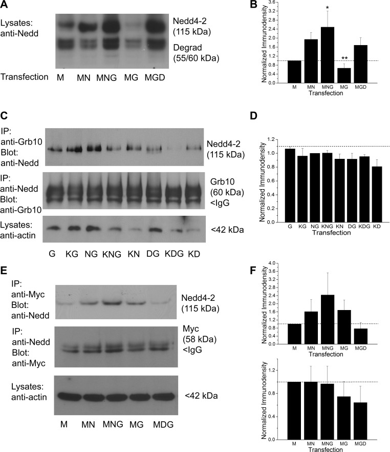
Fig. 7.
Grb10 adaptor protein enhances expression of Nedd4-2; Nedd4-2 forms a protein-protein interaction with Grb10 and with Kv1.3. A: protein separation strategy, Western blot, and transfection abbreviations as in Fig. 6. Nitrocellulose blots were probed with antiserum directed against Nedd4-2 (anti-Nedd, 1:2,000 dilution), Mr = 115 kDa. Note the increased expression of Nedd4-2 in the presence of Grb10 regardless of whether the catalytic site of the ligase was present or not (MNG vs. MGD). Note also a lower, doublet degradation product at 55/60 kDa, recognized by the manufacturer of the antibody. B: bar graph of the normalized immunodensity values of the collective protein bands for 4 experiments performed as in A; transfection conditions as noted. *P ≤ 0.05, **P ≤ 0.01, one-way ANOVA with SNK post hoc test. Dashed line, immunodensity value for Kv1.3 transfected condition alone. C: prepared lysates were either immunoprecipitated (IP) with Grb10 antibody (IP: anti-Grb10) and then blotted with Nedd4-2 (Blot: anti-Nedd, 1:2000), Mr = 115 kDa; or the reciprocal (IP: anti-Nedd/Blot: anti-Grb10, 1:800), Mr = 60 kDa. Input was also probed with anti-actin (1:800). D: bar graph of the normalized immunodensity values for three experiments performed for IP: anti-Nedd/Blot: anti-Grb10 as in C; notation and statistical analysis as above, not significantly different, P ≥ 0.05. E: prepared lysates were immunoprecipitated with myc antibody (IP: anti-Myc) and then blotted with Nedd4-2 (Blot: anti-Nedd, 1:2000), Mr = 115 kDa; or the reciprocal (IP:anti-Nedd/Blot: anti-Myc, 1:400), Mr = 58 kDa. Input was also probed with anti-actin (1:800). F: bar graph of the normalized immunodensity values for three experiments performed for each type of IP shown in E. Notation and statistical analyses as above, not significantly different, P ≥ 0.05.

