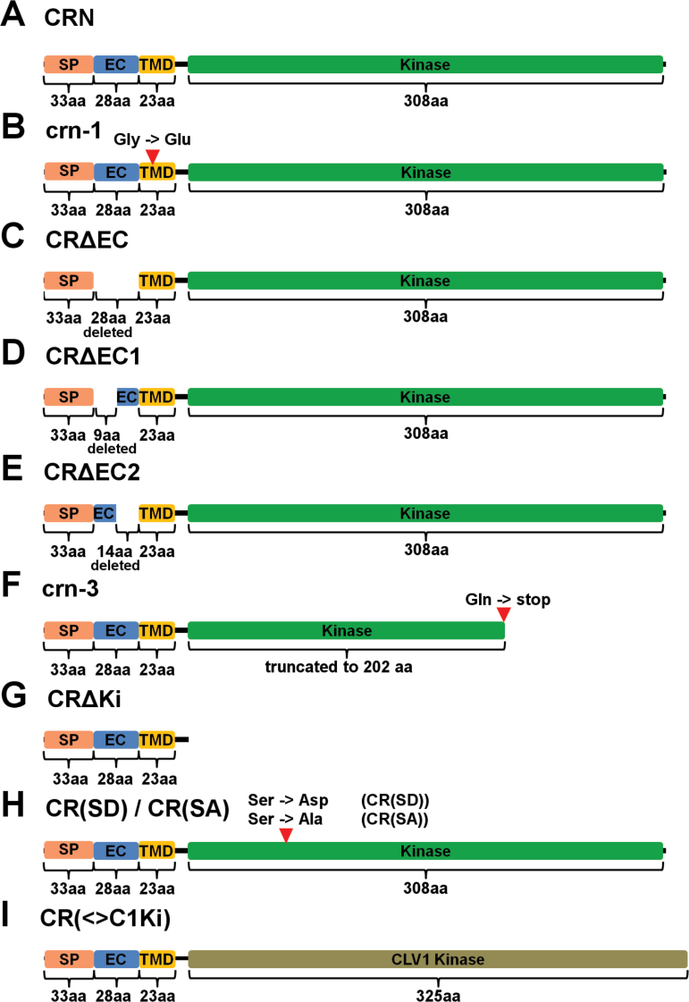
Fig. 1.
Schematic representations of the CRN protein variants. (A) CRN (wild-type); (B) crn-1; (C) CRΔEC; (D) CRΔEC1; (E) CRΔEC2; (F) crn-3; (G) CRΔKi; (H) CR(SD) and CR(SA); (I) CR (<>C1Ki). Light orange = signal peptide (SP); Blue = extracellular domain (EC); Yellow = transmembrane domain (TMD); Green = kinase domain; Olive = CLV1 kinase domain. aa = amino acids. The red arrowheads in (B), (F) and (H) indicate the positions of amino acid exchanges.

