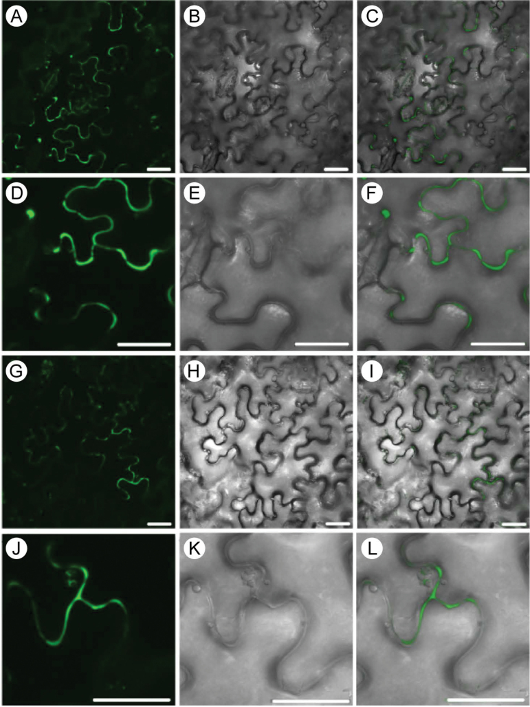
Fig. 5.
BiFC interaction between Serpin1 and SBT6.1. (A–F) Interaction between SBT6.1-nGFP and Serpin1-cGFP. (G–L) Interaction between SBT6.1-cGFP and Serpin1-nGFP. The subcellular localization was determined in the leaf epidermis of N. benthamiana (A–I). GFP fluorescence (A, D, G, and J), Nomarski differential interference contrast (DIC) (B, E, H, and K), and GFP/DIC overlapping images (C, F, I, and L). All images resulted from stacked confocal sections. Scale bars: 25 µm.

