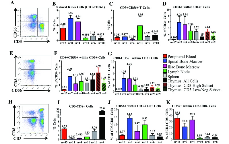
Figure 7.
Distribution of NK and CD56 across the hematopoietic and lymphoid organs of cynomolgus macaques. (A) Initially we defined NK cells as CD3–CD56+ cells. In each organ, we determined the presence of (B) NK cells and (C) CD3+CD56+ cells. We also examined (D) CD56 expression within CD3+ T cells. (E) Within T cells, we assessed the relationship between CD8 and CD56 expression. We examined both (F) coexpression of CD8 and CD56 and (G) expression of CD56 in the absence of CD8. (H) A population of CD3–CD8+ cells that has been suggested to have NK-like function in NHP was (I) observed and quantified in each tissue and (J) further examined for CD56 expression. (K) CD56+ expression within the CD3–CD8– population was evaluated as well. Data are given as mean ± SEM.

