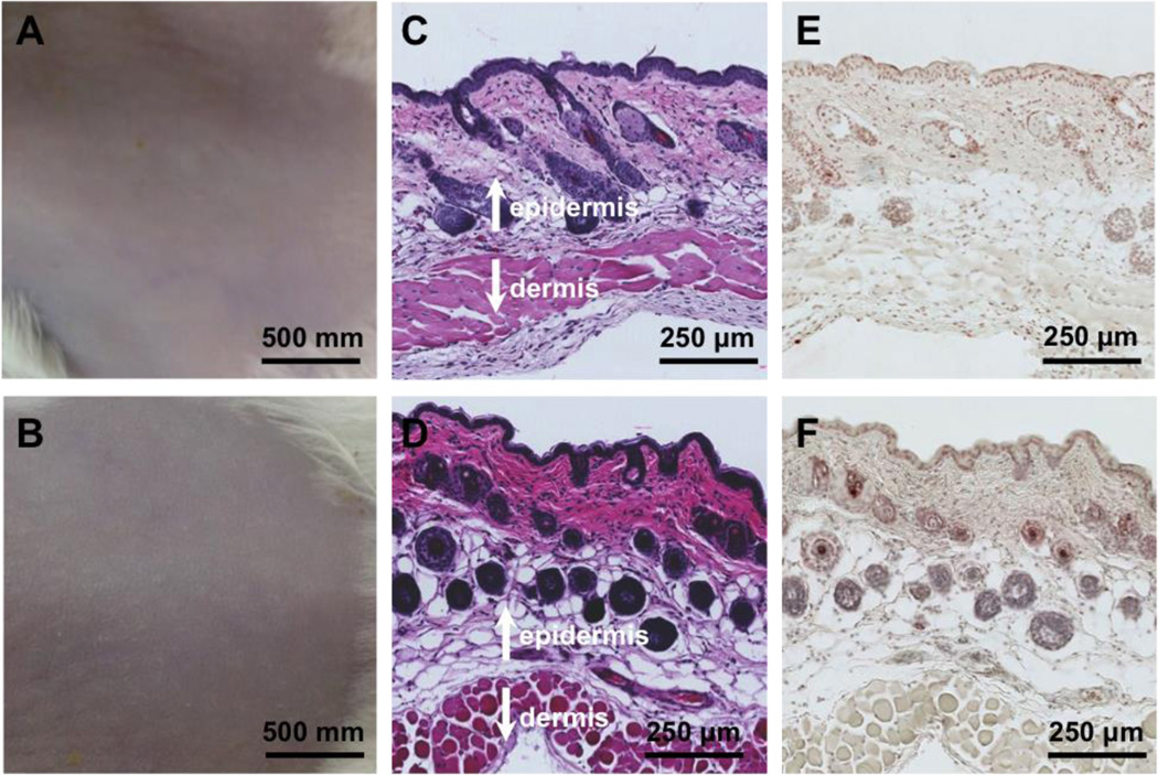
Figure 4.
Toxicity evaluation of the NP-gel using a mouse skin model. Mouse skin was treated with PBS buffer (A, C, and E) and NP-gel (B, D, and F), respectively. The samples were applied onto the shaved skin once a day for 7 days. Following the last treatment, the skin morphology of the two treatment groups was examined (A and B). The skin sections were further examined after H&E staining (C and D) and TUNEL staining (E and F).

